Log In


Hematite
A valid IMA mineral species - grandfathered
This page kindly sponsored by June Epp




About Hematite
Formula:
Fe2O3
Colour:
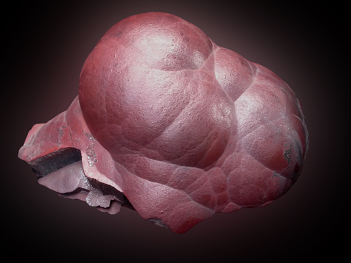
Steel-grey to black in crystals and massively crystalline ores, dull to bright "rust-red" in earthy, compact, fine-grained material.
Lustre:
Metallic, Sub-Metallic, Dull, Earthy
Hardness:
5 - 6
Specific Gravity:
5.26
Crystal System:
Trigonal
Member of:
Name:
Originally named about 300-325 BCE by Theophrastus from the Greek, "αιματίτις λίθος" ("aematitis lithos") for "blood stone". It is possibly the first mineral ever named ending with a "-ite" suffix. Translated in 79 by Pliny the Elder to haematites, "bloodlike", in allusion to the vivid red colour of the powder. The modern form evolved by authors frequently simplifying the spelling by excluding the "a", somewhat in parallel with other words originally utilising the root "haeme".
Dimorph of:
Hematite Group. The iron analogue of Corundum, Eskolaite, and Karelianite.
Hematite is rather variable in its appearance - it can be in reddish brown, ocherous masses, dark silvery-grey scaled masses, silvery-grey to black crystals, and dark-grey masses, to name a few. What they all have in common is a rust-red streak (except Ti-bearing varieties which have a black streak).
Black crystals may be confused with ilmenite.
NOTE: The 'hematite' used in jewelry, and often sold as magnetized items, is nothing of the sort and is an artificially created material, see Magnetic Hematite.
In an experimental volcanic gas condensation by Africano et al. (2002), it deposited in high fO2 conditions during cooling from ca. 800°C down to ca. 650°C.
 Visit gemdat.org for gemological information about Hematite.
Hematite is rather variable in its appearance - it can be in reddish brown, ocherous masses, dark silvery-grey scaled masses, silvery-grey to black crystals, and dark-grey masses, to name a few. What they all have in common is a rust-red streak (except Ti-bearing varieties which have a black streak).
Black crystals may be confused with ilmenite.
NOTE: The 'hematite' used in jewelry, and often sold as magnetized items, is nothing of the sort and is an artificially created material, see Magnetic Hematite.
In an experimental volcanic gas condensation by Africano et al. (2002), it deposited in high fO2 conditions during cooling from ca. 800°C down to ca. 650°C.
Visit gemdat.org for gemological information about Hematite.Unique Identifiers
Mindat ID:
1856
Long-form identifier:
mindat:1:1:1856:8
Similar Names
| Hemartite | A synonym of Bastnäsite-(Ce) | |
| Hematine | A synonym of 'Magnetic Hematite' | |
| Hematoide | A synonym of 'Ferruginous Quartz' | |
| Hepatite | A variety of Baryte | BaSO4 |
IMA Classification of Hematite
Approved, 'Grandfathered' (first described prior to 1959)
IMA Formula:
Fe3+2O3
Classification of Hematite
4.CB.05
4 : OXIDES (Hydroxides, V[5,6] vanadates, arsenites, antimonites, bismuthites, sulfites, selenites, tellurites, iodates)
C : Metal: Oxygen = 2: 3,3: 5, and similar
B : With medium-sized cations
4 : OXIDES (Hydroxides, V[5,6] vanadates, arsenites, antimonites, bismuthites, sulfites, selenites, tellurites, iodates)
C : Metal: Oxygen = 2: 3,3: 5, and similar
B : With medium-sized cations
Dana 7th ed.:
4.3.1.2
4.3.1.2
4 : SIMPLE OXIDES
3 : A2X3
4 : SIMPLE OXIDES
3 : A2X3
7.20.4
7 : Oxides and Hydroxides
20 : Oxides of Fe
7 : Oxides and Hydroxides
20 : Oxides of Fe
Mineral Symbols
As of 2021 there are now IMA–CNMNC approved mineral symbols (abbreviations) for each mineral species, useful for tables and diagrams.
Please only use the official IMA–CNMNC symbol. Older variants are listed for historical use only.
Please only use the official IMA–CNMNC symbol. Older variants are listed for historical use only.
| Symbol | Source | Reference for Standard |
|---|---|---|
| Hem | IMA–CNMNC | Warr, L.N. (2021). IMA–CNMNC approved mineral symbols. Mineralogical Magazine, 85(3), 291-320. doi:10.1180/mgm.2021.43 |
| Hem | Kretz (1983) | Kretz, R. (1983) Symbols of rock-forming minerals. American Mineralogist, 68, 277–279. |
| Hem | Siivolam & Schmid (2007) | Siivolam, J. and Schmid, R. (2007) Recommendations by the IUGS Subcommission on the Systematics of Metamorphic Rocks: List of mineral abbreviations. Web-version 01.02.07. IUGS Commission on the Systematics in Petrology. download |
| Hem | Whitney & Evans (2010) | Whitney, D.L. and Evans, B.W. (2010) Abbreviations for names of rock-forming minerals. American Mineralogist, 95, 185–187 doi:10.2138/am.2010.3371 |
| Hem | The Canadian Mineralogist (2019) | The Canadian Mineralogist (2019) The Canadian Mineralogist list of symbols for rock- and ore-forming minerals (December 30, 2019). download |
| Hem | Warr (2020) | Warr, L.N. (2020) Recommended abbreviations for the names of clay minerals and associated phases. Clay Minerals, 55, 261–264 doi:10.1180/clm.2020.30 |
Pronunciation of Hematite
Pronunciation:
| Play | Recorded by | Country |
|---|---|---|
| Jolyon Ralph | United Kingdom |
Physical Properties of Hematite
Metallic, Sub-Metallic, Dull, Earthy
Transparency:
Opaque
Colour:
Steel-grey to black in crystals and massively crystalline ores, dull to bright "rust-red" in earthy, compact, fine-grained material.
Comment:
See Rossman, G. R. (1996) for cause of red colour.
Streak:
Reddish brown ("rust-red"); blackish when Ti-bearing
Hardness:
5 - 6 on Mohs scale
Hardness:
VHN100=1000 - 1100 kg/mm2 - Vickers
Hardness Data:
Measured
Tenacity:
Brittle
Cleavage:
None Observed
Parting:
Partings on {0001} and {1011} due to twinning. Unique cubic parting in masses and grains at Franklin Mine, Franklin, NJ.
Fracture:
Irregular/Uneven, Sub-Conchoidal
Comment:
Elastic in thin lamellae
Density:
5.26 g/cm3 (Measured) 5.255 g/cm3 (Calculated)
Optical Data of Hematite
Type:
Uniaxial (-)
RI values:
nω = 3.15 - 3.22 nε = 2.87 - 2.94
Max. Birefringence:
δ = 0.280
Based on recorded range of RI values above.
Based on recorded range of RI values above.
Interference Colours:
The colours simulate birefringence patterns seen in thin section under crossed polars. They do not take into account mineral colouration or opacity.
Michel-Levy Bar The default colours simulate the birefringence range for a 30 µm thin-section thickness. Adjust the slider to simulate a different thickness.
Grain Simulation You can rotate the grain simulation to show how this range might look as you rotated a sample under crossed polars. Each grain retains its interference colour (retardation) while its brightness falls to black at extinction and reaches a maximum between extinction positions.
The colours simulate birefringence patterns seen in thin section under crossed polars. They do not take into account mineral colouration or opacity.
Michel-Levy Bar The default colours simulate the birefringence range for a 30 µm thin-section thickness. Adjust the slider to simulate a different thickness.
Grain Simulation You can rotate the grain simulation to show how this range might look as you rotated a sample under crossed polars. Each grain retains its interference colour (retardation) while its brightness falls to black at extinction and reaches a maximum between extinction positions.
Surface Relief:
Very High (positive)
Relative to Canada balsam mounting medium (n ≈ 1.537).
Relative to Canada balsam mounting medium (n ≈ 1.537).
This shows the grain boundary and Becke line effect under plane-polarised
light, based on the contrast between this mineral's average refractive
index and the mounting medium. It does not take into account mineral
colouration.
In focus
Interference Figure:
This shows the idealized uniaxial interference figure - the conoscopic
(convergent-light, Bertrand-lens-in) view, for a grain cut with the optic axis
centred and vertical. The coloured rings are isochromatics, computed with the
same physics as the Michel-Lévy bar above; the dark cross is the isogyre.
For a genuinely uniaxial mineral viewed this way, that cross stays perfectly stationary if you rotate the stage - unlike a biaxial mineral, where it splits apart on rotation. That invariance is itself the standard diagnostic test for telling uniaxial and biaxial minerals apart at the microscope.
For a genuinely uniaxial mineral viewed this way, that cross stays perfectly stationary if you rotate the stage - unlike a biaxial mineral, where it splits apart on rotation. That invariance is itself the standard diagnostic test for telling uniaxial and biaxial minerals apart at the microscope.
Anisotropism:
Distinct
Reflectivity:
| Wavelength | R1 (%) | R2 (%) | imR1 (%) | imR2 (%) |
|---|---|---|---|---|
| 400nm | 26.8% | 30.5% | 12.2% | 15.6% |
| 420nm | 28.5% | 31.8% | 13.9% | 17.0% |
| 440nm | 28.9% | 32.1% | 14.3% | 17.3% |
| 460nm | 28.2% | 31.9% | 13.6% | 17.0% |
| 470nm | 28.1% | 31.7% | 13.4% | 16.8% |
| 480nm | 27.9% | 31.6% | 13.3% | 16.7% |
| 500nm | 27.5% | 31.3% | 12.9% | 16.3% |
| 520nm | 27.2% | 30.5% | 12.6% | 15.6% |
| 540nm | 26.7% | 30.1% | 12.2% | 15.3% |
| 546nm | 26.4% | 30.0% | 12.0% | 15.1% |
| 560nm | 26.1% | 29.8% | 11.8% | 15.0% |
| 580nm | 25.5% | 29.3% | 11.3% | 14.6% |
| 600nm | 24.8% | 28.6% | 10.8% | 14.0% |
| 620nm | 24.1% | 27.7% | 10.3% | 13.2% |
| 640nm | 23.6% | 26.7% | 9.9% | 12.4% |
| 650nm | 23.3% | 26.3% | 9.7% | 12.0% |
| 660nm | 23.0% | 25.9% | 9.5% | 11.7% |
| 680nm | 22.6% | 25.3% | 9.2% | 11.2% |
| 700nm | 22.3% | 25.1% | 9.0% | 11.1% |
Graph shows reflectance levels at different wavelengths (in nm). Peak reflectance is 32.1%.
R1 shown in black, R2 shown in red, imR1 shown in green, imR2 shown in blue
Colour in reflected light:
White to greyish white with bluish tint
Internal Reflections:
Red
Pleochroism:
Weak
Comments:
O = brownish red
E = yellowish red
E = yellowish red
Chemistry of Hematite
Mindat Formula:
Fe2O3
Elements listed:
Common Impurities:
Ti,Al,Mn,H2O
Age distribution
Recorded ages:
Mesozoic : 236 ± 7 Ma to 145 ± 4 Ma - based on 6 recorded ages.
Sample ages:
| Sample ID | Recorded age | Geologic Time | Dating method |
|---|---|---|---|
| 1 | 145 ± 4 Ma | Late Jurassic | U-Th/He |
| 2 | 183 ± 13 Ma | Early Jurassic | (U-Th)/He |
| 3 | 236 ± 7 Ma | Late/Upper Triassic | U-Th/He |
Sample references:
Chemical Analysis
Oxide wt%:
| 1 | 2 | 3 | |
|---|---|---|---|
| Fe2O3 | 87.0 % | 86.95 % | |
| MnO2 | 2.5 % | 2.93 % | |
| SiO2 | 5.0 % | 8.20 % | |
| Al2O3 | 2.0 % | 0.73 % | 0.12 % |
| Loss | 3.5 % | 1.19 % | |
| TiO2 | 5.69 % | ||
| MnO | 0.16 % | ||
| FeO | 88.91 % | ||
| MgO | 0.47 % | ||
| Total: | 100 % | 100 % | 95.35 % |
Empirical formulas:
| Sample ID | Empirical Formula |
|---|---|
| 3 | (Fe3+,Al)1.774(Fe2+, Mg, Mn)0.113Ti0.113O3 |
Sample references:
| ID | Locality | Reference | Notes |
|---|---|---|---|
| 1 | Bihain, Vielsalm, Luxembourg, Wallonia, Belgium | ||
| 2 | Colanhan, Lierneux, Liège, Wallonia, Belgium | ||
| 3 | Dypingdal serpentine-magnesite deposit, Snarum, Modum, Buskerud, Norway | Microprobe, Fe2+ |
Crystallography of Hematite
Crystal System:
Trigonal
Class (H-M):
3m(32/m) - Hexagonal Scalenohedral
Space Group:
R3c
Cell Parameters:
a = 5.038(2) Å, c = 13.772(12) Å
Ratio:
a:c = 1 : 2.734
Unit Cell V:
302.72 ų (Calculated from Unit Cell)
Z:
6
Morphology:
Crystals generally thick to thin tabular {0001}, rarely prismatic [0001] or scalenohedral; also rarely rhombohedral {1011}, producing pseudo-cubic crystals. Often found in sub-parallel growths on {0001} or as rosettes ("iron roses.") Sometimes in micaceous to platy masses. May be compact columnar or fibrous masses, sometimes radiating, or in reniform masses with a smooth fracture ("kidney ore"), and botryoidal and stalactic. Frequently in earthy masses, also granular, friable to compact, concretionary and oolitic.
Twinning:
Penetration twins on {0001}, or with {1010} as a composition plane. Frequently exhibits a lamellar twinning on {1011} in polished section.
Crystallographic forms of Hematite
Crystal Atlas:
Image Loading
Click on an icon to view

Hematite no.319 - {001} - Goldschmidt (1913-1926)
Hematite no.331 - {0.1.10} - Goldschmidt (1913-1926)
Hematite no.337 - {102} - Goldschmidt (1913-1926)
3d models and HTML5 code kindly provided by
www.smorf.nl.
Toggle
Edge Lines | Miller Indices | Axes
Transparency
Opaque | Translucent | Transparent
View
Along a-axis | Along b-axis | Along c-axis | Start rotation | Stop rotation
Edge Lines | Miller Indices | Axes
Transparency
Opaque | Translucent | Transparent
View
Along a-axis | Along b-axis | Along c-axis | Start rotation | Stop rotation
Crystal Structure
Load
Unit Cell | Unit Cell Packed
2x2x2 | 3x3x3 | 4x4x4
Unit Cell | Unit Cell Packed
2x2x2 | 3x3x3 | 4x4x4
Show
Big Balls | Small Balls | Just Balls | Spacefill
Polyhedra Off | Si Polyhedra | All Polyhedra
Remove metal-metal sticks
Big Balls | Small Balls | Just Balls | Spacefill
Polyhedra Off | Si Polyhedra | All Polyhedra
Remove metal-metal sticks
Display Options
Black Background | White Background
Perspective On | Perspective Off
2D | Stereo | Red-Blue | Red-Cyan
Black Background | White Background
Perspective On | Perspective Off
2D | Stereo | Red-Blue | Red-Cyan
View
CIF File Best | x | y | z | a | b | c
CIF File Best | x | y | z | a | b | c
Rotation
Stop | Start
Stop | Start
Labels
Console Off | On | Grey | Yellow
Console Off | On | Grey | Yellow
Data courtesy of the American Mineralogist Crystal Structure Database. Click on an AMCSD ID to view structure
| ID | Species | Reference | Link | Year | Locality | Pressure (GPa) | Temp (K) |
|---|---|---|---|---|---|---|---|
| 0017808 | Hematite | Maslen E N, Streltsov V A, Streltsova N R, Ishizawa N (1994) Synchrotron X-ray study of the electron density in alpha-Fe2O3 Acta Crystallographica B50 435-441 |  | 1994 | synthetic | 0 | 293 |
| 0017807 | Hematite | Maslen E N, Streltsov V A, Streltsova N R, Ishizawa N (1994) Synchrotron X-ray study of the electron density in alpha-Fe2O3 Acta Crystallographica B50 435-441 | | 1994 | synthetic | 0 | 293 |
| 0017806 | Hematite | Maslen E N, Streltsov V A, Streltsova N R, Ishizawa N (1994) Synchrotron X-ray study of the electron density in alpha-Fe2O3 Acta Crystallographica B50 435-441 | | 1994 | synthetic | 0 | 293 |
| 0017731 | Hematite | Finger L W, Hazen R M (1980) Crystal structure and isothermal compression of Fe2O3, Cr2O3, and V2O3 to 50 kbars Journal of Applied Physics 51 5362-5367 | 1980 | synthetic | 0.0001 | 293 | |
| 0000143 | Hematite | Blake R L, Hessevick R E, Zoltai T, Finger L W (1966) Refinement of the hematite structure American Mineralogist 51 123-129 |  | 1966 | Elba, Italy | 0 | 293 |
| 0017732 | Hematite | Finger L W, Hazen R M (1980) Crystal structure and isothermal compression of Fe2O3, Cr2O3, and V2O3 to 50 kbars Journal of Applied Physics 51 5362-5367 | 1980 | synthetic | 1.54 | 293 | |
| 0017733 | Hematite | Finger L W, Hazen R M (1980) Crystal structure and isothermal compression of Fe2O3, Cr2O3, and V2O3 to 50 kbars Journal of Applied Physics 51 5362-5367 | 1980 | synthetic | 3.14 | 293 | |
| 0017734 | Hematite | Finger L W, Hazen R M (1980) Crystal structure and isothermal compression of Fe2O3, Cr2O3, and V2O3 to 50 kbars Journal of Applied Physics 51 5362-5367 | 1980 | synthetic | 4.16 | 293 | |
| 0017735 | Hematite | Finger L W, Hazen R M (1980) Crystal structure and isothermal compression of Fe2O3, Cr2O3, and V2O3 to 50 kbars Journal of Applied Physics 51 5362-5367 | 1980 | synthetic | 4.39 | 293 | |
| 0017736 | Hematite | Finger L W, Hazen R M (1980) Crystal structure and isothermal compression of Fe2O3, Cr2O3, and V2O3 to 50 kbars Journal of Applied Physics 51 5362-5367 | 1980 | synthetic | 5.24 | 293 | |
| 0018106 | Hematite | Pauling L, Hendricks S (1925) The Structure of Hematite Journal of the American Chemical Society 47 781-790 | 1925 | 0 | 293 | ||
| 0014076 | Hematite | Pauling L, Hendricks S B (1925) Crystal structures of hematite and corundum Journal of the American Chemical Society 47 781-790 | 1925 | 0 | 293 | ||
| 0002228 | Hematite | Gualtieri A, Venturelli P (1999) In situ study of the goethite-hematite phase transformation by real time synchrotron powder diffraction American Mineralogist 84 895-904 | | 1999 | 0 | 586 | |
| 0002229 | Hematite | Gualtieri A, Venturelli P (1999) In situ study of the goethite-hematite phase transformation by real time synchrotron powder diffraction American Mineralogist 84 895-904 | | 1999 | 0 | 790 | |
| 0002230 | Hematite | Gualtieri A, Venturelli P (1999) In situ study of the goethite-hematite phase transformation by real time synchrotron powder diffraction American Mineralogist 84 895-904 | | 1999 | 0 | 1052 |
CIF Raw Data - click here to close
Epitaxial Relationships of Hematite
Epitaxial Minerals:
| 'Pseudobrookite' | Fe3+2Ti4+O5 |
| 'Rutile' | TiO2 |
Epitaxy Comments:
Examples of rutile epitaxial on hematite are widespread. Dramatic specimens have been found at Novo Horizonte, Brazil.
Pseudobrookite on hematite with pseudobrookite {121}[210] parallel to hematite {0001}[1100].
Pseudobrookite on hematite with pseudobrookite {121}[210] parallel to hematite {0001}[1100].
X-Ray Powder Diffraction
Loading XRD data...
Data courtesy of RRUFF project at University of Arizona, used with permission.
Powder Diffraction Data:
| d-spacing | Intensity |
|---|---|
| 3.68 Å | (30) |
| 2.70 Å | (100) |
| 2.52 Å | (70) |
| 2.21 Å | (20) |
| 1.84 Å | (40) |
| 1.69 Å | (50) |
| 1.49 Å | (30) |
| 1.45 Å | (30) |
Geological Environment
Paragenetic Mode(s):
Geological Setting:
Large ore bodies of hematite are usually of sedimentary origin; also found in high-grade ore bodies in metamorphic rocks due to contact metasomatism, and occasionally as a sublimate on igneous extrusive rocks ("lavas") as a result of volcanic activity. It is also usually the cause of red soils all over the planet.
Synonyms of Hematite
Other Language Names for Hematite
Basque:Hematite
Catalan:Hematites
Croatian:Hematit
Czech:Hematit
Dutch:Hematiet
Esperanto:Hematito
Estonian:Hematiit
Finnish:Hematiitti
French:Hématite
Fer oligiste
Fer oxydé rouge
Hematite rouge
Fer oxydé (in part)
Titane oxydé ferrifère (in part)
Fer oligiste
Fer oxydé rouge
Hematite rouge
Fer oxydé (in part)
Titane oxydé ferrifère (in part)
Galician:Hematita
Saint-Christophe-en-Oisans, Grenoble, Isère, Auvergne-Rhône-Alpes, France