Log In


Bakerite
A variety of Datolite
This page is currently not sponsored. Click here to sponsor this page.


About Bakerite
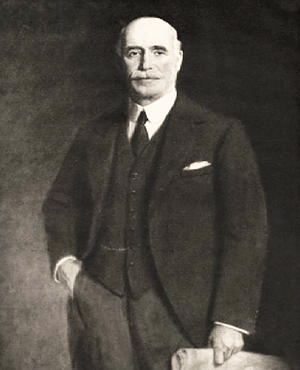
Richard Charles Baker
Formula:
Ca4(H5B5Si3O20)
Colour:
Colourless, White
Lustre:
Vitreous, Dull
Hardness:
4½
Specific Gravity:
2.88
Crystal System:
Monoclinic
Name:
Named in 1903 by William Brantingham Giles in honor of Richard Charles Baker (1858 Islington, London, England, UK - 1937 California, USA), who discovered the species, then president of the Borax Consolidated Company (formerly the Borax Company Limited) of San Bernardino County, California, USA, and for whom Baker, California, USA is also named. (Earlier, Richard C. Baker was also president of the Pacific Coast Borax Company, a company probably related to his later business interests.)
A microcrystalline, boron-rich variety of datolite. Occurs in various genetic environments among them diabase spilites.
Unique Identifiers
Mindat ID:
490 (as Bakerite)
1340 (as Datolite)
1340 (as Datolite)
Long-form identifier:
mindat:1:1:490:5 (as Bakerite)
mindat:1:1:1340:2 (as Datolite)
mindat:1:1:1340:2 (as Datolite)
Similar Names
IMA Classification of Bakerite
Discredited
Approval history:
Discredited: IMA 16-A. See Hålenius et al. (2016) and Bačík et al. (2017).
Classification of Bakerite
54.2.1b.1
54 : NESOSILICATES Borosilicates and Some Beryllosilicates
2 : Borosilicates and Some Beryllosilicates with B in [4] coordination
54 : NESOSILICATES Borosilicates and Some Beryllosilicates
2 : Borosilicates and Some Beryllosilicates with B in [4] coordination
17.5.13
17 : Silicates Containing other Anions
5 : Borosilicates
17 : Silicates Containing other Anions
5 : Borosilicates
Physical Properties of Bakerite
Vitreous, Dull
Transparency:
Translucent
Colour:
Colourless, White
Hardness:
4½ on Mohs scale
Density:
2.88 g/cm3 (Measured) 2.94 g/cm3 (Calculated)
Optical Data of Bakerite
Type:
Biaxial (-)
RI values:
nα = 1.624 nβ = 1.635 nγ = 1.654
2V:
Measured: 87° to 88°
Birefringence:
Moderate
Max. Birefringence:
δ = 0.030
Based on recorded range of RI values above.
Based on recorded range of RI values above.
Interference Colours:
The colours simulate birefringence patterns seen in thin section under crossed polars. They do not take into account mineral colouration or opacity.
Michel-Levy Bar The default colours simulate the birefringence range for a 30 µm thin-section thickness. Adjust the slider to simulate a different thickness.
Grain Simulation You can rotate the grain simulation to show how this range might look as you rotated a sample under crossed polars. Each grain retains its interference colour (retardation) while its brightness falls to black at extinction and reaches a maximum between extinction positions.
The colours simulate birefringence patterns seen in thin section under crossed polars. They do not take into account mineral colouration or opacity.
Michel-Levy Bar The default colours simulate the birefringence range for a 30 µm thin-section thickness. Adjust the slider to simulate a different thickness.
Grain Simulation You can rotate the grain simulation to show how this range might look as you rotated a sample under crossed polars. Each grain retains its interference colour (retardation) while its brightness falls to black at extinction and reaches a maximum between extinction positions.
Surface Relief:
High (positive)
Relative to Canada balsam mounting medium (n ≈ 1.537).
Relative to Canada balsam mounting medium (n ≈ 1.537).
This shows the grain boundary and Becke line effect under plane-polarised
light, based on the contrast between this mineral's average refractive
index and the mounting medium. It does not take into account mineral
colouration.
In focus
Interference Figure:
This shows the idealized biaxial acute bisectrix (Bxa) interference figure
- the conoscopic view for a grain cut perpendicular to the acute bisectrix, using
this mineral's 2V. The two small white dots mark the melatopes - the points
where the two optic axes emerge - and are shown only when they fall within the
field of view. The coloured bands are isochromatics, and the dark bands are
isogyres.
Rotate the stage: at 0°/90° the isogyres form a cross through the melatopes; at 45° they pull apart into curved hyperbolas. That splitting on rotation - absent in a uniaxial figure - is the standard diagnostic test for telling biaxial minerals from uniaxial ones. If 2V is large, the melatopes may fall outside the field of view, as they often do at the microscope too.
Rotate the stage: at 0°/90° the isogyres form a cross through the melatopes; at 45° they pull apart into curved hyperbolas. That splitting on rotation - absent in a uniaxial figure - is the standard diagnostic test for telling biaxial minerals from uniaxial ones. If 2V is large, the melatopes may fall outside the field of view, as they often do at the microscope too.
Dispersion:
weak
Rio Gavottino, Casarza Ligure, Genoa, Liguria, Italy